Endorectal ultrasound examination
Endorectal ultrasound examination is ultrasound examination, which is performed by high frequency probe through rectum.
Endorectal ultrasound examination is one of the most informative methods of detection for extent of rectal cancer invasion, lymph nodes involvement, topographical diagnosis of anal abscesses and fistulas, perfect imagining of prostate and possibility to fine the smallest pathology in investigated tissue.
Intestinal wall, spaces surrounding the rectum, extend of different disease (cancers, polyps, fistulas, affected lymph nodes) are investigated by 3D images. One of the most important advantages of this method is the possibility of prostate and parirectal fat biopsy controlled by endorectal ultrasound.
Endorectal ultrasound examination can be performed only after adequate preparation. Method of examination The exam usually is carried out in the lateral decubitus position. The examination is performed in accurate continuity: primarily anal canal, sphincter system, layers of intestinal wall and only after that prostate.
Endorectal ultrasound examination is not radiation method, that’s why if dynamic observation is necessary it can by performed several times.
Endorectal ultrasound examination in case of rectal cancer
Traditional trans-abdominal diagnostic of rectal cancer often is complicated because of artifacts conditioned by depth disposition of rectum, acoustical dark of pubic bone and gas in rectum. Endorectal ultrasound is one of the most informative examinations of rectal cancer invasion depth and lymph nodes involvement.
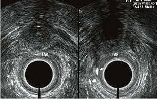
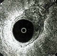
Normally, anal canal has three-layer structure (mucosa with submucosa, internal sphincter and external sphincter). Rectal wall also has three-layer structure (mucosa with submucosa, muscular and serous layers). The most important criterion of tumor’s invasive growth is defection in normal structure of intestinal layers. Local thickness of any intestinal wall can be the feature of invasive growth. Cancer’s infiltration of perirectal fat has also spatial signs.
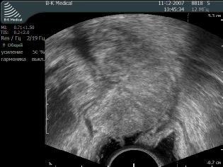
Diagnostic effectiveness of endorectal ultrasound is especially high for early cancer (T1-T2). In this case cancer borders and nearby tissue infiltration are identified easily. Sensitivity of this method for early tumor and locally advanced tumors is 91%, peculiarity – 89%, that is more than the same index of CT and MRI.
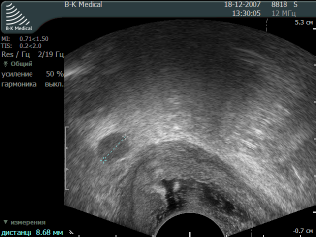
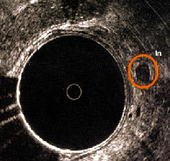
Metastasis in lymph nodes looks like bigger than 0,3cm circle formations in perirectal fat. Overstaging is possible in case, when nodes are inflamed (they can look almost like nodes with metastasis).
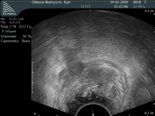
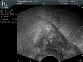
Rectal cancer T2
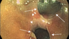
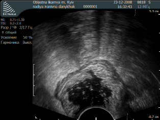
Anal fistula
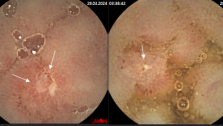
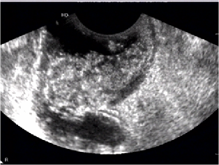
Rectal cancer T4
Metastasis in lymph node of perirectal fat
Rectal cancer T4N1
Rectal cancer T4NO
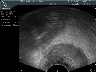
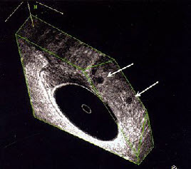
Rectal cancer T4
Rectal cancer T3N1
Rectal cancer T3NO
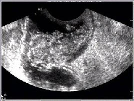
Rectal cancer T2NO
Lymph nodes with metastasis
Rectal cancer T3NO
Lymph nodes with metastasis
Lymph nodes with metastasis
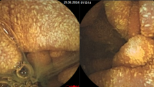
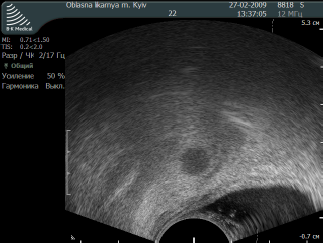
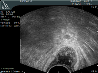
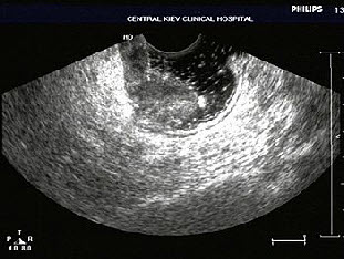
Malignant polyp of rectum
Read more: Preparation for endorectal ultrasound examination