On 09.04.23, Dr Milianovska A.O. performed video capsule endoscopy
On 09.04.23, Dr Milianovska A.O. performed video capsule endoscopy on a patient born in 1993, who was referred for further examination by the attending physician from another region. The anamnesis showed that in 2019, against the background of acute intestinal obstruction, the patient underwent a small bowel resection with a benign large polyp, which caused the obstruction. A month ago, the situation repeated itself. The patient was again operated on at the place of residence against the background of acute intestinal obstruction. But this time, the histology was different from the previous one, it was a highly differentiated adenocarcinoma in a glandular polyp of the small intestine. After the surgery, the patient underwent a CT scan of the peritoneum with contrast at the place of residence. The CT scan showed no evidence of a small bowel neoplasm. In addition, a gastroscopy was performed. According to the patient, no one had previously recommended an additional bowel examination.
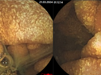
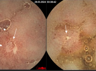
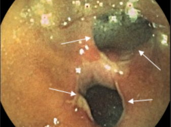
The video capsule endoscopy clearly visualised a p/o scar in the jejunum. Distal to the scar, an exophytic neoplasm, a polyp, 10-15 mm in diameter, is visualised. The approximate location of the neoplasm is c/3 of the jejunum. In addition, there are single polyps of 3-5 mm in size, type 0Іs in the stomach.


